Purified anti-Pax-6 Antibody (Previously Covance catalog# PRB-278P)

Pricing & Availability
Clone
Poly19013 (See other available formats)
Regulatory Status
RUO
Other Names
Paired box protein Pax-6, oculorhombin, aniridia type II protein, Sey, Protein eyeless, PRB-278P-100
Previously
Covance Catalog# PRB-278P
Isotype
Rabbit Polyclonal IgG
Ave. Rating
Submit a Review
Product Citations
publications
1-Poly19013_PURE_Pax-6_Antibody_1_091420
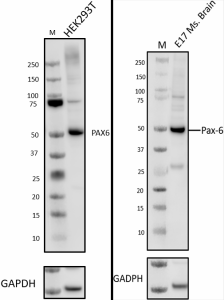
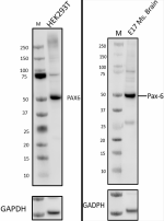
Whole cell extracts (15µg total protein) from 293T (human), (left panel) or E17 Ms. brain (mouse brain at day 17 of development), (right panel) were resolved by 4-12% Bis-Tris gel electrophoresis, transferred to PVDF membrane and probed with 1.0 µg/mL (1:2000 dilution) of purified Pax-6 rabbit antibody (clone Poly19013) for 2 hr at RT. Proteins were visualized by chemiluminescence detection using HRP Donkey anti-rabbit IgG Antibody (Cat. No. 406401, 1:3000 dilution). Direct-Blot™ HRP anti-GAPDH antibody (Cat. No. 607904) was used as a loading control at 1:25000 dilution (lower). Lane M:Molecular weight marker.
  • 1-Poly19013_PURE_Pax-6_Antibody_1_091420
    Whole cell extracts (15µg total protein) from 293T (human), (left panel) or E17 Ms. brain (mouse brain at day 17 of development), (right panel) were resolved by 4-12% Bis-Tris gel electrophoresis, transferred to PVDF membrane and probed with 1.0 µg/mL (1:2000 dilution) of purified Pax-6 rabbit antibody (clone Poly19013) for 2 hr at RT. Proteins were visualized by chemiluminescence detection using HRP Donkey anti-rabbit IgG Antibody (Cat. No. 406401, 1:3000 dilution). Direct-Blot™ HRP anti-GAPDH antibody (Cat. No. 607904) was used as a loading control at 1:25000 dilution (lower). Lane M:Molecular weight marker.
  • 2-Poly19013_PURE_Pax-6_Antibody_2_091420
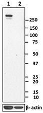
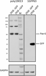
    Whole cell extracts (15 µg total protein) from either untransfected 293E cells or transfected with GFP were resolved by 4-12% Bis-Tris gel electrophoresis, transferred to PVDF membrane and probed with 1.0 µg/mL (1:2000 dilution) of purified Pax-6 rabbit antibody (clone Poly19013) (panel A) or purified anti-GFP antibody (clone 1GFP63, Cat. No. 668206) for 2 hr at RT. Proteins were visualized by chemiluminescence detection using HRP Donkey anti-rabbit IgG Antibody (Cat. No. 406401, 1:3000 dilution) or HRP Goat anti-mouse IgG Antibody (Cat. No. 405306, 1:3000 dilution). Direct-Blot™ HRP anti-GAPDH antibody (Cat. No. 607904) was used as a loading control at 1:25000 dilution (lower).Lane M:Molecular weight marker.
  • 3_Purified_Pax-6_Antibody_IHC_2_021315
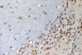
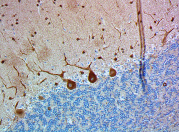
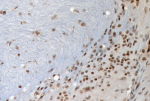
    IHC staining of purified anti-Pax-6 antibody (clone Poly19013) on formalin-fixed paraffin-embedded mouse brain tissue. Following antigen retrieval using Sodium Citrate H.I.E.R., the tissue was incubated with 20 µg/ml of the primary antibody for 60 minutes at room temperature. BioLegend's Ultra-Streptavidin (USA) HRP kit (Multi-Species, DAB, Cat. No. 929901) was used for detection followed by hematoxylin counterstaining, according to the protocol provided. The image was captured with a 40X objective.
Cat # Size Price Quantity Check Availability Save
901302 25 µL 109€
Check Availability


Need larger quantities of this item?
Request Bulk Quote
901301 100 µL 331€
Check Availability


Need larger quantities of this item?
Request Bulk Quote
Description

Pax6 is a transcription factor present during embryonic development. The encoded protein contains two different binding sites that are known to bind DNA and function as a regulator of gene transcription. It is a key regulatory gene of eye and brain development. Within the brain, the protein is involved in development of specialized cells that process smell. Pax-6 acts as a critical gene for the development of eyes and other sensory organs, certain neural and epidermal tissues as well as other homologous structures, usually derived from ectodermal tissues. Pax6 serves as a regulator in the coordination and pattern formation required for differentiation and proliferation to successfully take place, ensuring that the processes of neurogenesis and oculogenesis are carried out successfully. As a transcription factor, Pax6 acts at the molecular level in the signaling and formation of the central nervous system. The characteristic paired DNA binding domain of Pax6 utilizes two DNA-binding domains, the paired domain (PD), and the paired-type homeodomain (HD). These domains function separately. An example of this lies in HD’s regulatory involvement in the formation of the lens and retina throughout oculogenesis contrasted by the molecular mechanisms of control exhibited on the patterns of neurogenesis in brain development by PD. The HD and PD domains act in close coordination, giving Pax6 its multifunctional nature in directing molecular signaling in formation of the CNS.

The vertebrate PAX6 locus encodes at least three different protein isoforms, these being the canonical PAX6, PAX6(5a), and PAX6(ΔPD). The canonical PAX6 protein contains an N-terminal paired domain, connected by a linker region to a paired-type homeodomain, and a proline/serine/threonine (P/S/T)-rich C-terminal domain. The paired domain and paired-type homeodomain each have DNA binding activities, while the P/S/T-rich domain possesses a transactivation function. PAX6(5a) is a product of the alternatively spliced exon 5a resulting in a 14 residue insertion in the paired domain which alters the specificity of this DNA binding activity. The nucleotide sequence corresponding to the linker region encodes a set of three alternative translation start codons from which the third PAX6 isoform originates. Collectively known as the PAX6(ΔPD) or pairedless isoforms, these three gene products all lack a paired domain. The pairedless proteins possess molecular weights of 43, 33, or 32kDa, depending on the particular start codon used. PAX6 transactivation function is attributed to the variable length C-terminal P/S/T-rich domain which stretches to 153 residues in human and mouse proteins.

Product Details
Technical Data Sheet (pdf)

Product Details

Verified Reactivity
Human, Mouse, Rat
Antibody Type
Polyclonal
Host Species
Rabbit
Immunogen
This antibody was generated against the peptide (QVPGSEPDMSQYWPRLQ) derived from the C-terminus of the mouse Pax-6 protein.
Formulation
Phosphate-buffered solution + 0.03% Thimerosal.
Preparation
The antibody was purified by affinity chromatography.
Concentration
2 mg/mL
Storage & Handling
The antibody solution should be stored undiluted between 2°C and 8°C. Please note the storage condition for this antibody has been changed from -20°C to between 2°C and 8°C. You can also check your vial or your CoA to find the most accurate storage condition for this antibody.
Application

WB, IHC-P - Quality tested
IHC-F - Reported in the literature, not verified in house

Recommended Usage

Each lot of this antibody is quality control tested by Western blotting and formalin-fixed paraffin-embedded immunohistochemical staining of brain tissue. For Western blotting, the suggested use of this reagent is 1.0 µg/mL (1:2000). For immunohistochemistry, a dilution of 1:50 - 1:100 is suggested. It is recommended that the reagent be titrated for optimal performance for each application.

Application Notes

This antibody weakly reacts with rat. It has also shown reactivity with GFP and is therefore not recommended for use in GFP-expressing systems (as determined by in-house testing).

Pax-6 has two isoforms in human and mice at 46.6 and 48.2 Kd. This antibody recognizes both. The sequence is highly conserved among Pax-6 of various species. The antibody was subsequently purified on a Protein A column and is useful in studying brain, neuronal and olfactory development in higher eukaryotes.

This clone is not recommended for ChIP (Chromatin Immunoprecipitation) assays (as determined by in-house testing).

Application References
  1. Tang K, et al. 2012. Development. 139:1630. (IF) PubMed
  2. Bandah D, et al. 2007. Invest. Ophthalmol. Vis. Sci. 48:2503. (WB) PubMed
  3. Marquardt T, et al. 2001. Cell. 105:43.
  4. Yamamoto Y, Jeffery WR. 2000. Science. 289:631
  5. Ashery-Padan R, et al. 2000. Genes Dev. 14:2701.
  6. Osumi N, et al. 1997. Development. 124:2961.
  7. Koroma BM, et al. 1997. Invest. Ophthalmol Vis Sci. 38:108.
  8. Davis JA, Reed RR. 1996. J Neurosci. 16:5082. (IHC)
  9. Quadrato G, et al. Nature. 545:48. (IF) PubMed
Product Citations
  1. Subramanian L, et al. 2017. Nat Commun. 8:14167. PubMed
  2. Schmid B, et al. 2021. Stem Cell Research. 52:102180. PubMed
  3. Ma L, et al. 2021. Stem Cell Research. 53:102305. PubMed
  4. Li J, et al. 2021. Stem Cell Research. 53:102376. PubMed
  5. Martens YA, et al. 2021. STAR Protocols. 2(2):100571. PubMed
  6. Hwang SH, et al. 2021. eLife. 0.416666666666667. PubMed
  7. Tristan CA, et al. 2020. bioRxiv. . PubMed
  8. Takahashi K, et al. 2021. PLoS Genet. 17:e1009587. PubMed
  9. Ziffra RS, et al. 2021. Nature. 598:205. PubMed
  10. Schroeter MS, et al. 2022. MRS Bull. 47:530. PubMed
  11. Ahmad Z, et al. 2015. PLoS One. 10: 0144597. PubMed
  12. Ramzy A, et al. 2018. Endocrinology. 159:83. PubMed
  13. Scior A, et al. 2018. EMBO J. 37:282. PubMed
  14. Wilson CS, et al. 2019. JCI Insight. 5. PubMed
  15. Nazir FH, et al. 2018. Neurochem Int. 121:38. PubMed
  16. Bashford AL, et al. 2019. J Pathol. 248:396. PubMed
  17. Del Toro D et al. 2017. Cell. 169(4):621-635 . PubMed
  18. Popova G, et al. 2021. Cell Stem Cell. 28:2153. PubMed
  19. Muralidharan H, et al. 2022. J Neurosci. 42:2149. PubMed
  20. Jabali A, et al. 2022. EMBO Rep. 23:e54027. PubMed
  21. Jia X, et al. 2022. Sci Adv. 8:eabo7112. PubMed
  22. Chen X, et al. 2023. Neuron. 111:30. PubMed
  23. Sterling NA, et al. 2023. Nat Commun. 14:82. PubMed
  24. Kokorudz C, et al. 2022. Cells. 12: . PubMed
  25. Zhong Z, et al. 2022. Bio Protoc. 12: . PubMed
  26. Xue W, et al. 2023. iScience. 26:105898. PubMed
  27. Cheng Q, et al. 2023. Mol Brain. 16:24. PubMed
  28. Zhong DJ, et al. 2021. FASEB J. 35:e21842. PubMed
  29. Le Floch P, et al. 2022. Adv Mater. 34:e2106829. PubMed
  30. Lorenz C, et al. 2022. Stem Cell Res. 61:102742. PubMed
  31. Brozko N, et al. 2022. Front Neuroanat. 16:838567. PubMed
  32. Gallo RA, et al. 2022. Transl Vis Sci Technol. 11:4. PubMed
  33. Habib K, et al. 2022. Mol Cells. 45:588. PubMed
  34. Wang S, et al. 2022. J Neurosci. 42:7031. PubMed
  35. Tarazi S, et al. 2022. Cell. 185:3290. PubMed
  36. Wang L, et al. 2022. Proc Natl Acad Sci U S A. 119:e2206147119. PubMed
  37. , et al. 2022. Nature. 609:1021. PubMed
  38. Whye D, et al. 2023. Curr Protoc. 3:e641. PubMed
  39. Dong Z, et al. 2023. J Biol Chem. 299:103020. PubMed
  40. Loan A, et al. 2023. iScience. 26:106093. PubMed
  41. Li B, et al. 2023. Cell Discov. 9:27. PubMed
  42. Guo CJ, et al. 2023. Mol Ther Nucleic Acids. 31:763. PubMed
  43. Xu A, et al. 2023. Nat Commun. 14:1694. PubMed
  44. Eigenhuis KN, et al. 2023. Front Cell Neurosci. 17:1114420. PubMed
  45. Cappuccio G, et al. 2023. Front Mol Biosci. 10:1181965. PubMed
  46. Rybak-Wolf A, et al. 2023. Nat Microbiol. . PubMed
  47. Merkert S, et al. 2021. Stem Cell Research. 52:102208. PubMed
  48. Gao X, et al. 2020. Stem Cell Research. 48:101986. PubMed
  49. Patmore DM, et al. 2020. Developmental Cell. 54(4):455-470.e5. PubMed
  50. Viais R, et al. 2021. Elife. 10:. PubMed
  51. Avilés EC, et al. 2022. Cell Rep. 38:110307. PubMed
  52. Notaras M, et al. 2021. Mol Psychiatry. Online ahead of print. PubMed
  53. Taniguchi-Ikeda M, et al. 2021. iScience. 24:103140. PubMed
  54. Tian E, et al. 2016. Cell Rep. 16: 781-792. PubMed
  55. Small K, et al. 2015. Ophthalmology. 13: 681-685. PubMed
  56. Bershteyn M, et al. 2017. Cell Stem Cell. 20:435. PubMed
  57. Panagiotakos G, et al. 2019. Elife. 8:e51037. PubMed
  58. Peter CJ, et al. 2019. Nat Commun. 10:4112. PubMed
  59. Bovio PP, et al. 2019. Mol Neurobiol. 56:4273. PubMed
  60. Ing-Esteves S, et al. 2018. J Neurosci. 38:2713. PubMed
  61. Sun X, et al. 2021. Stem Cell Research. 52:102230. PubMed
  62. Kafarnik C, et al. 2020. Stem Cells Dev. 425:29. PubMed
  63. Sood S, et al. 2020. Proc Natl Acad Sci U S A. 22331:117. PubMed
  64. Junqueira Alves C, et al. 2021. Nat Commun. 12:6019. PubMed
  65. Moura D, et al. 2016. Sci Rep. 6:28532. PubMed
  66. Bonnefont J, et al. 2019. Neuron. 103:1096. PubMed
  67. Hickmott JW, et al. 2018. Gene Ther. 25:524. PubMed
  68. Liu J et al. 2017. Cell stem cell. 21(5):635-649 . PubMed
  69. Liu X, et al. 2021. Cell Prolif. 54:e13124. PubMed
  70. Kalusa M, et al. 2021. Front Neuroanat. 15:678385. PubMed
  71. Gregory JA, et al. 2020. Cells. 9:00. PubMed
  72. de Leeuw VC, et al. 2020. Cell Reprogram. 1.125. PubMed
  73. Ucuncu E, et al. 2020. Nat Commun. 4.685416667. PubMed
  74. Girskis KM, et al. 2021. Neuron. 109:3239. PubMed
  75. Singer RA, et al. 2020. Cell Metabolism. 30(6):1091-1106. PubMed
  76. Saito K, et al. 2019. Cell Rep. 29:1555. PubMed
  77. Slot E, et al. 2020. Stem Cell Res. 101745:44. PubMed
  78. Sun WR, et al. 2020. Hum Mol Genet. . PubMed
  79. Freude KK, et al. 2020. World J Stem Cells. 1171:12. PubMed
  80. LaForce GR, et al. 2022. Neuron. 110:1340. PubMed
  81. Bandah D, et al. 2007. Invest Ophthalmol Vis Sci. 48:2503-2509. PubMed
  82. Pataskar A, et al. 2016. EMBO J. 35: 24 - 45. PubMed
  83. Broix L, et al. 2016. Nat Genet. 48:1349-1358. PubMed
  84. Chen Y, et al. 2019. Methods Mol Biol. 1919:59:00. PubMed
  85. Moore D, et al. 2019. Stem Cells Int. 2019:8710180. PubMed
  86. Komabayashi-Suzuki M, et al. 2019. Cell Rep. 29:1113. PubMed
  87. Selvadurai HJ, et al. 2020. Cell Reports. 31(2):107511.. PubMed
  88. Shao W, et al. 2020. Nature. 580:106. PubMed
  89. Janssens S, et al. 2018. mSystems. 3. PubMed
  90. Tripathy R, et al. 2018. Neuron. 100:1354. PubMed
  91. Wang H, et al. 2019. Cell Rep. 27:2335. PubMed
  92. Tristan CA, et al. 2022. Nat Protoc. Online ahead of print. PubMed
  93. Ezan J, et al. 2021. Sci Rep. 9106:11. PubMed
  94. Nakashima H, et al. 2021. Cell Reports. 35(7):109124. PubMed
  95. Lardelli R, et al. 2017. Nat Genet. 10.1038/ng.3762. PubMed
  96. Taverna E, et al. 2016. Sci Rep. 6:21206. PubMed
  97. Ahn LY, et al. 2021. J Neurosci Res. 99:110. PubMed
  98. Birkhoff JC, et al. 2020. Hum Mol Genet. 29:2535. PubMed
  99. Pereiro X, et al. 2020. Cells. 9:00. PubMed
  100. Kim TW, et al. 2021. Cell Stem Cell. 28(2):343-355.e5. PubMed
  101. Simsa R, et al. 2021. PLoS One. 16:e0245685. PubMed
  102. Li M, et al. 2021. Nat Commun. 12:420. PubMed
  103. Craenen K, et al. 2020. Reprod Toxicol. 91:59. PubMed
  104. Walker H, et al. 2021. Int J Mol Sci. 22:. PubMed
  105. Hou S, et al. 2021. Cereb Cortex. 31:4730. PubMed
  106. Delépine C, et al. 2016. Hum Mol Genet. 25: 146 - 157. PubMed
  107. Xu J, et al. 2016. Sci Transl Med. 8: 333ra48. PubMed
  108. Bonney S, et al. 2016. J Neurosci. 36: 7786 - 7801. PubMed
  109. Pei S, et al. 2016. Sci Rep. 6:33822. PubMed
  110. M Rodriguez, S Noctor, E Muñoz 2016. PLoS One. 11:e0167063. PubMed
  111. Boland M, et al. 2017. Brain. 10.1093/brain/aww357. PubMed
  112. Matsumura K, et al. 2020. Nat Commun. 11:859. PubMed
  113. Lee EJ, et al. 2020. Cell Reports. 28(3):712-722.e3.. PubMed
  114. Roy A, et al. 2020. Acta Neuropathologica Communications. 8(1):54.. PubMed
  115. Shafa M, et al. 2018. Front Med (Lausanne). 9:1816. PubMed
  116. Zhang X, et al. 2019. Protein Cell. 10:649. PubMed
  117. Postel M, et al. 2019. BMC Bioinformatics. 20:470. PubMed
  118. Ma L, et al. 2022. iScience. 25:105172. PubMed
  119. Li N, et al. 2021. Stem Cell Research. 53:102322. PubMed
  120. Gao X, et al. 2021. Stem Cell Research. 53:102271. PubMed
  121. Schinzel F, et al. 2021. Journal of Comparative Neurology. 529(11):3032-3045. PubMed
  122. Xiao D, et al. 2020. Sci Adv. 6:eaaz5858. PubMed
  123. Retallack H, et al. 2016. Proc Natl Acad Sci U S A. 113(50):14408-14413. PubMed
  124. Simonin Y, et al. 2016. EBioMedicine. 12:161-169. PubMed
  125. Otani T, et al. 2016. Cell Stem Cell. 18: 467-480. PubMed
  126. Tomita H, et al. 2020. Cell Reports. 30(1):215-228.e5.. PubMed
  127. Plaza Reyes A, et al. 2020. Nat Commun. 11:1609. PubMed
  128. Wang Q, et al. 2017. Curr Biol. 27:1791. PubMed
  129. Todd L, et al. 2017. Glia. 65:1640. PubMed
  130. Lu HC, et al. 2017. Nat Genet. 49:527. PubMed
  131. Dhumale P, et al. 2018. PLoS One. 9:1544. PubMed
  132. Chen X, et al. 2022. PLoS Biol. 20:e3001868. PubMed
  133. Bosze B, et al. 2020. J Neurosci. 40:1501. PubMed
  134. Eze UC, et al. 2021. Nature Neuroscience. 24(4):584-594. PubMed
  135. Chen M, et al. 2021. Stem Cell Research. 51:102166. PubMed
  136. Marczenke M, et al. 2021. Development. 148:. PubMed
  137. Zhu X, et al. 2022. Sci Adv. 8:eabl7263. PubMed
  138. Harkin L, et al. 2016. Cereb Cortex. 10.1093/cercor/bhw394. PubMed
  139. Fregeac J, et al. 2020. Mol Autism. 11:22. PubMed
  140. Li L, et al. 2019. Cell Rep. 28:698. PubMed
  141. Omer Javed A, et al. 2018. Cell Rep. 25:368. PubMed
  142. Sun D, et al. 2017. PLoS One. 12(6):e0179047. PubMed
  143. Chau KF, et al. 2018. Elife. 19:E663. PubMed
  144. Andres LM, et al. 2017. ACS Chem Biol. 12:2030. PubMed
  145. Ibañez Rodriguez MP et al. 2018. The Journal of comparative neurology. 526(15):2462-2481 . PubMed
  146. Pollen AA, et al. 2019. Cell. 176:743. PubMed
  147. Cliff TS et al. 2017. Cell stem cell. 21(4):502-516 . PubMed
  148. Urciuolo A, et al. 2020. Nat Biomed Eng. 4:901. PubMed
  149. Ramme AP, et al. 2021. Stem Cell Research. 53:102327. PubMed
  150. Zhang L, et al. 2020. Cell Stem Cell. 27(5):784-797.e11. PubMed
  151. Jeong D, et al. 2020. Cell Rep. 33:108257. PubMed
  152. Haldipur P, et al. 2021. Acta Neuropathol. 142:761. PubMed
  153. Li J, et al. 2021. Neural Plast. 2021:4894881. PubMed
  154. Schembs L, et al. 2022. Cell Rep. 39:110811. PubMed
  155. Soubannier V, et al. 2022. Cells. 11:. PubMed
  156. Manos JD, et al. 2022. iScience. 25:103658. PubMed
  157. Han X, et al. 2021. J Neurosci. Online ahead of print. PubMed
  158. Breuss M, et al. 2017. Hum Mol Genet. 10.1093/hmg/ddw383. PubMed
  159. Paquet D, et al. 2016. Nature. 533: 125-129. PubMed
  160. Ramme AP, et al. 2020. Stem Cell Research. 41:101615.. PubMed
  161. Popovitchenko T, et al. 2020. Nat Commun. 11:1674. PubMed
  162. Velasco S, et al. 2019. Nature. 570:523. PubMed
  163. Forrest MP, et al. 2017. Cell Stem Cell. 1.086805556. PubMed
  164. Lavado A et al. 2018. Dev Cell. 47(5):576-591 . PubMed
  165. Docampo–Seara A, et al. 2018. Brain Struct Funct. 223:3593. PubMed
  166. Langer LF, et al. 2019. Elife. 8. PubMed
  167. Hoshino A, et al. 2017. Dev Cell. 43:763. PubMed
  168. Luisier R, et al. 2018. Nat Commun. 8:6458. PubMed
  169. Ren R, et al. 2017. Cell Res. 10.1038/cr.2017.18. PubMed
  170. Ao Z, et al. 2020. Anal Chem. 4630:92. PubMed
  171. Li YP, et al. 2021. STAR Protoc. 2:100438. PubMed
  172. Clark AE, et al. 2020. J Virol. 95:e02024. PubMed
  173. Chui A, et al. 2020. Development. . PubMed
  174. Chao J, et al. 2022. iScience. 25:104391. PubMed
  175. Okamoto M, et al. 2016. Nat Commun. 7: 11349. PubMed
  176. Hickmott J, et al. 2016. Mol Ther Methods Clin Dev. 3:16051. PubMed
  177. Zhu L, et al. 2016. J Cell Biol. 215: 187 - 202. PubMed
  178. Wörsdörfer P, et al. 2019. Sci Rep. 9:15663. PubMed
  179. Pandey PR, et al. 2019. Int J Mol Sci. 0.936805556. PubMed
  180. Mirjalili Mohanna SZ, et al. 2020. Mol Ther Methods Clin Dev. 17:478. PubMed
  181. Yin WC, et al. 2019. Dev Cell. 48:167. PubMed
  182. Goodson NB, et al. 2018. Dev Biol. 434:149. PubMed
  183. Ren X, et al. 2019. Cell Rep. 26:3643. PubMed
  184. Beattie R et al. 2017. Neuron. 94(3):517-533 . PubMed
  185. Parween S, et al. 2018. Front Cell Neurosci. 0.746527778. PubMed
  186. Padmanabhan Nair V, et al. 2021. Cell Stem Cell. . PubMed
  187. Chui A, et al. 2020. Development. 147:00:00. PubMed
  188. Quintana-Urzainqui I, et al. 2020. Development. 147:00:00. PubMed
  189. Gey M, et al. 2016. J Neurosci. 36: 9057 - 9069. PubMed
  190. Sancho-Martinez I, et al. 2016. Nat Commun. 7:10743. PubMed
  191. Shu P, et al. 2019. J Biol Chem. 294:3444. PubMed
  192. Mao X, et al. 2019. Stem Cell Reports. 13:747. PubMed
  193. Lindsay CD, et al. 2019. Acta Biomater. 95:225. PubMed
  194. Shafa M, et al. 2019. Int J Mol Sci. 0.95. PubMed
  195. Gabriel E et al. 2017. Cell stem cell. 20(3):397-406 . PubMed
  196. Homman‐Ludiye J et al. 2018. The Journal of comparative neurology. 526(17):2870-2883 . PubMed
  197. Xu D, et al. 2018. PLoS Biol. 16:e2006613. PubMed
  198. Zahr SK et al. 2018. Neuron. 97(3):520-537 . PubMed
  199. Kim YJ, et al. 2018. Neuron. 100:1180. PubMed
  200. Tu J et al. 2018. Stem cell research. 28:29-32 . PubMed
  201. Mills T, et al. 2017. PLoS One.. 10.1371/journal.pone.0176905. PubMed
  202. Kim HM, et al. 2020. Cell Prolif. :e12965. PubMed
  203. Usman A, et al. 2021. Stem Cell Research. 55:102488. PubMed
  204. Kaufman ML, et al. 2021. Development. . PubMed
  205. Romero DM, et al. 2022. Nat Commun. 13:2746. PubMed
  206. Fischer M, et al. 2021. Alcohol Clin Exp Res. 45:979. PubMed
  207. Prince LM, et al. 2021. Food Chem Toxicol. 152:112178. PubMed
  208. Behesti H, et al. 2021. Elife. 10:. PubMed
  209. Garreta E, et al. 2016. Biomaterials. 98:64-78. PubMed
  210. Robson JP, et al. 2019. PLoS One. 14:e0210665. PubMed
  211. Remez LA, et al. 2017. Dev Biol. 432:140. PubMed
  212. Klein T et al. 2019. Stem cell research. 1724:56:00 . PubMed
  213. Sano N et al. 2017. Frontiers in cellular neuroscience. 0.54375 . PubMed
  214. Chai G, et al. 2020. Neuron. 109(2):241-256.e9. PubMed
  215. Yu Q, et al. 2021. eLife. 10:00. PubMed
  216. Miyashita H, et al. 2017. Sci Rep. 7:43557. PubMed
  217. Marinopoulou E, et al. 2021. iScience. 24:103198. PubMed
  218. Tachibana N, et al. 2016. J Neurosci. 36: 9454 - 9471. PubMed
  219. Lin H, et al. 2016. Nature. 531:323-328. PubMed
  220. Wang W, et al. 2016. Nat Commun. 7:10936. PubMed
  221. Bando Y, et al. 2016. Cereb Cortex. 26: 106 - 117. PubMed
  222. Cheng S, et al. 2019. J Neurosci. 39:2195. PubMed
  223. Uzquiano A, et al. 2019. Cell Rep. 28:1596. PubMed
  224. Docampo–Seara A, et al. 2019. Brain Struct Funct. 224:2325. PubMed
  225. Lim Y, et al. 2019. Sci Rep. 9:226. PubMed
  226. Nakano Y et al. 2018. Cell. 174(3):536-548 . PubMed
  227. Zhu Y, et al. 2017. Lab Chip. 10.1039/c7lc00682a. PubMed
  228. Metzger JJ, et al. 2022. Cell Rep Methods. 2:100297. PubMed
  229. Li M, et al. 2021. Signal Transduct Target Ther. 6:05. PubMed
  230. Kitajima R, et al. 2020. Stem Cell Res. 101749:44. PubMed
  231. Inak G, et al. 2021. Nat Commun. 12:1929. PubMed
  232. Liang D, et al. 2021. Nat Neurosci. 24:941. PubMed
  233. Vajhøj C, et al. 2021. PLoS One. 16:e0261536. PubMed
  234. Nieto-Estévez V, et al. 2022. Front Mol Neurosci. 14:815808. PubMed
  235. Nieto-Estévez V, et al. 2022. Front Mol Neurosci. 15:889922. PubMed
  236. Tang K, et al. 2012. Development. 139:1630-1639. PubMed
  237. He S, et al. 2016. Development. 143: 1937 - 1947. PubMed
  238. Lajko M, et al. 2016. PLoS One. 11:e0166886. PubMed
  239. Hasenpusch–Theil K, et al. 2020. eLife. 9:e58162. PubMed
  240. Bendriem RM, et al. 2020. eLife. 8:e49376.. PubMed
  241. Wu X, et al. 2018. Cell. 172:423. PubMed
  242. Shibata S et al. 2018. Cell reports. 25(6):1668-1679 . PubMed
  243. Thomas Klein et al. 2018. Stem cell research. 33:171-174 . PubMed
  244. Ward JM, et al. 2019. Cell Rep. 26:1189. PubMed
  245. Wang S, et al. 2019. Protein Cell. 10:1. PubMed
  246. Watanabe Y, et al. 2018. Development. 145. PubMed
  247. Wu Z, et al. 2019. Neuron. 101:635. PubMed
  248. Miesfeld JB, et al. 2018. Sci Rep. 8:10195. PubMed
  249. Meng Y, et al. 2021. Front Cell Dev Biol. 9:702462. PubMed
  250. Mathieu C, et al. 2021. MBio. 12:e0079921. PubMed
  251. Lin L, et al. 2020. Neuron. 107:1180. PubMed
  252. Aldinger KA, et al. 2021. Nat Neurosci. 24:1163. PubMed
  253. Carpentieri JA, et al. 2022. Nat Commun. 13:16. PubMed
  254. C Tao, X Zhang, 2016. Cell Rep. 17:1832-1844. PubMed
  255. Damiani D, et al. 2016. Nat Commun. 7:13509. PubMed
  256. Toda T, et al. 2016. Sci Rep. 6:29578. PubMed
  257. Anda F, et al. 2016. Nat Neurosci. 10.1038/nn.4328. PubMed
  258. Martínez-Cerdeño V, et al. 2016. Cereb Cortex. 26: 374 - 383. PubMed
  259. Yao H, et al. 2020. Sci Rep. 10:17445. PubMed
  260. Ding W, et al. 2019. J Neurosci. 39:1994. PubMed
  261. Gao ML, et al. 2020. Front Cell Dev Biol. 8:128. PubMed
  262. Gao F et al. 2019. Mol Cell. 75(5):891-904 . PubMed
  263. Sanchez–Guardado L, et al. 2019. Elife. 8:e46675. PubMed
  264. Kurabayashi N, et al. 2018. Development. 145. PubMed
  265. Kawaue T, et al. 2019. Nat Commun. 10:2780. PubMed
  266. Nowakowski TJ, et al. 2018. Nat Neurosci. 21:1784. PubMed
  267. Baizabal JM et al. 2018. Neuron. 98(5):945-962 . PubMed
  268. Arber C, et al. 2021. Cell Reports. 34(2):108615. PubMed
  269. Kweon H, et al. 2021. Cell Reports. 34(8):108780. PubMed
  270. Bowles KR, et al. 2021. Cell. 184(17):4547-4563.e17. PubMed
  271. Campbell WA, et al. 2021. Glia. 69:1515. PubMed
  272. Li Z, et al. 2020. ACS Infect Dis. 6:2616. PubMed
  273. Too LK, et al. 2021. Retina. 41:1986. PubMed
  274. Wind M, et al. 2021. Curr Protoc. 1:e244. PubMed
  275. Conrad E, et al. 2016. Am J Physiol Endocrinol Metab. 310: E91 - E102. PubMed
  276. Wang L, Hou S, Han Y 2016. Nat Neurosci. 19: 888-896. PubMed
  277. Abdullah A, et al. 2016. Stem Cell Reports. 7:69-79. PubMed
  278. Yan P, et al. 2020. Cell Rep. 32:107870. PubMed
  279. Thomas Klein et al. 2018. Stem cell research. 31:222-226 . PubMed
  280. Han S, et al. 2018. Development. 145:. PubMed
  281. Palencia–Campos A, et al. 2017. Hum Mol Genet. 26:4556. PubMed
  282. Karzbrun E, et al. 2018. Nat Phys. 14:515. PubMed
  283. Mochizuki T, et al. 2017. Development. 144:708. PubMed
  284. Alzu'bi A et al. 2019. Journal of anatomy. 235(3):555-568 . PubMed
  285. Korecki AJ, et al. 2021. Gene Ther. 28:351. PubMed
  286. Besusso D, et al. 2020. Stem Cell Res. 102016:49. PubMed
  287. Jia N, et al. 2021. Stem Cell Research. 53:102367. PubMed
  288. Somatilaka BN, et al. 2020. Developmental Cell. 54(6):710-726.e8. PubMed
  289. Arnst N, et al. 2021. Cells. 10:. PubMed
  290. Bockaj I, et al. 2021. PLoS Genet. 17:e1009868. PubMed
  291. Cugola F, et al. 2016. Nature. 534: 267-271. PubMed
  292. Radonjić N, et al. 2016. Cereb Cortex. 26: 131 - 143. PubMed
  293. Todd L, et al. 2018. Stem Cells. 36:392. PubMed
  294. Segarra M, et al. 2018. Science. 361:eaao2861. PubMed
  295. Jin J, et al. 2019. PLoS One. 14:e0219362. PubMed
  296. Alexander JM et al. 2019. Elife. 8 pii: e41769. PubMed
  297. Zurkirchen L, et al. 2019. Nat Commun. 10:2192. PubMed
  298. Diacou R et al. 2018. Cell reports. 25(9):2510-2523 . PubMed
  299. Tang Y et al. 2018. Cell reports. 24(5):1355-1362 . PubMed
  300. Krach F, et al. 2022. Nat Commun. 13:6797. PubMed
  301. Tsai MH, et al. 2020. Acta Neuropathol Commun. 0.406944444. PubMed
  302. Cederquist GY, et al. 2020. Cell Stem Cell. 27(1):35-49.e6. PubMed
  303. Haag D, et al. 2021. Cancer Cell. 39(3):407-422.e13. PubMed
  304. Bannier-Hlaout M, et al. 2021. Cell Stem Cell. 28(7):1221-1232.e7. PubMed
  305. Tolomeo AM, et al. 2021. Front Cell Neurosci. 602888:15. PubMed
  306. Chen Y, et al. 2021. Nat Methods. 18:528. PubMed
  307. Penna E, et al. 2021. Cereb Cortex Commun. 2:tgab053. PubMed
  308. Kaufman ML, et al. 2021. Development. 148:. PubMed
  309. Yao X, et al. 2022. Nat Commun. 13:3883. PubMed
  310. Yang Y, et al. 2022. Front Bioeng Biotechnol. 10:907159. PubMed
  311. Verheyen A, et al. 2015. PLoS One. 10: 0146127. PubMed
  312. Takeo Y, et al. 2016. Sci Rep. 6: 25180. PubMed
  313. Plaisted W, et al. 2016. PLoS One. 11: 0157620. PubMed
  314. Feng J, et al. 2016. Cereb Cortex. 26: 3323 - 3334. PubMed
  315. Zhang Y et al. 2018. Cell stem cell. 23(4):516-529 . PubMed
  316. Le Dréau G, et al. 2018. Elife. 7:e7267. PubMed
  317. Broix L, et al. 2018. Hum Mol Genet. 27:224. PubMed
  318. Dominguez Gonzalez B, et al. 2018. Hum Mol Genet. 27:2154. PubMed
  319. Ooi J, et al. 2019. Cell Rep. 26:2494. PubMed
  320. Hu XL, et al. 2017. Neuron. 95:309. PubMed
  321. Chang CH, et al. 2019. Dev Cell. 48:184. PubMed
  322. Ferdaos N, et al. 2022. PLoS One. 17:e0278147. PubMed
  323. Patel A, et al. 2020. Development. dev193649. PubMed
  324. Žagare A, et al. 2021. STAR Protoc. 2:100524. PubMed
  325. Gao X, et al. 2021. Stem Cell Research. 53:102388. PubMed
  326. Li Z, et al. 2020. Emerg Microbes Infect. 2.044444444. PubMed
  327. Hayashi R, et al. 2017. Nat Protoc. 10.1038/nprot.2017.007. PubMed
  328. Barra JM, et al. 2021. Xenotransplantation. 28:e12706. PubMed
  329. Zhang YF, et al. 2022. Front Cell Neurosci. 16:866020. PubMed
  330. Zhang XZ, et al. 2022. Front Cell Neurosci. 16:888152. PubMed
  331. Torsahakul C, et al. 2022. PLoS One. 17:e0263141. PubMed
  332. Klein JA, et al. 2022. Front Cell Neurosci. 15:794675. PubMed
  333. Wiley L, et al. 2016. Sci Rep. 6:30742. PubMed
  334. Laan L, et al. 2020. Clin Epigenetics. 12:09. PubMed
  335. Liu Z, et al. 2019. Cell Rep. 27:3832. PubMed
  336. Roy A et al. 2019. Elife. 8 pii: e45961. PubMed
  337. Cargnin F, et al. 2018. Neuron. 100:1083. PubMed
RRID
AB_2749901 (BioLegend Cat. No. 901302)
AB_2565003 (BioLegend Cat. No. 901301)

Antigen Details

Cell Type
Neural Stem Cells
Biology Area
Cell Biology, Neuroscience, Neuroscience Cell Markers, Signal Transduction, Stem Cells, Synaptic Biology, Transcription Factors
Molecular Family
Nuclear Markers
Gene ID
5080 View all products for this Gene ID 18508 View all products for this Gene ID 25509 View all products for this Gene ID
UniProt
View information about Pax-6 on UniProt.org

Related FAQs

There are no FAQs for this product.

Other Formats

View All Pax-6 Reagents Request Custom Conjugation
Description Clone Applications
Purified anti-Pax-6 Poly19013 WB,IHC-P,IHC-F
Go To Top Version: 9    Revision Date: 08/24/2021

For Research Use Only. Not for diagnostic or therapeutic use.


This product is supplied subject to the terms and conditions, including the limited license, located at www.biolegend.com/terms) ("Terms") and may be used only as provided in the Terms. Without limiting the foregoing, BioLegend products may not be used for any Commercial Purpose as defined in the Terms, resold in any form, used in manufacturing, or reverse engineered, sequenced, or otherwise studied or used to learn its design or composition without express written approval of BioLegend. Regardless of the information given in this document, user is solely responsible for determining any license requirements necessary for user’s intended use and assumes all risk and liability arising from use of the product. BioLegend is not responsible for patent infringement or any other risks or liabilities whatsoever resulting from the use of its products.


BioLegend, the BioLegend logo, and all other trademarks are property of BioLegend, Inc. or their respective owners, and all rights are reserved.


8999 BioLegend Way, San Diego, CA 92121 www.biolegend.com
Toll-Free Phone: 1-877-Bio-Legend (246-5343) Phone: (858) 768-5800 Fax: (877) 455-9587

This data display is provided for general comparisons between formats.
Your actual data may vary due to variations in samples, target cells, instruments and their settings, staining conditions, and other factors.
If you need assistance with selecting the best format contact our expert technical support team.
ProductsHere

Login / Register
Remember me
Forgot your password? Reset password?
Create an Account