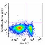
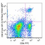
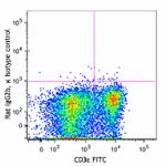
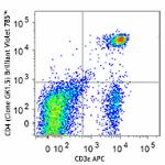
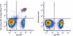
FITC anti-mouse CD4 Antibody

Pricing & Availability
Clone
GK1.5 (See other available formats)
Regulatory Status
RUO
Other Names
L3T4, T4
Isotype
Rat IgG2b, κ
Ave. Rating
Submit a Review
Product Citations
publications
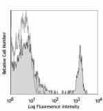
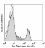
GK1.5_FITC_102110
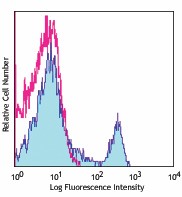
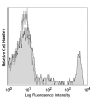
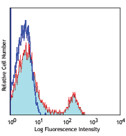
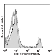
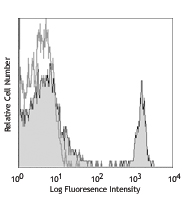
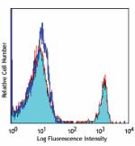
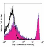
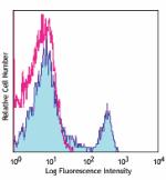
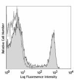
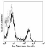
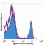
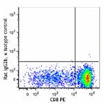
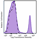
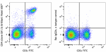
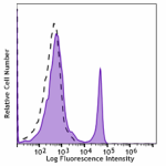
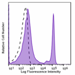
C57BL/6 mouse splenocytes were stained with CD4 (clone GK1.5) FITC (filled histogram) or rat IgG2b, κ FITC isotype control (open histogram).
  • GK1.5_FITC_102110
    C57BL/6 mouse splenocytes were stained with CD4 (clone GK1.5) FITC (filled histogram) or rat IgG2b, κ FITC isotype control (open histogram).
Compare all formats See FITC spectral data
Cat # Size Price Quantity Check Availability Save
100405 50 µg 24€
Check Availability


Need larger quantities of this item?
Request Bulk Quote
100406 500 µg 80€
Check Availability


Need larger quantities of this item?
Request Bulk Quote
Description

CD4 is a 55 kD protein also known as L3T4 or T4. It is a member of the Ig superfamily, primarily expressed on most thymocytes, a subset of T cells, and weakly on macrophages and dendritic cells. It acts as a coreceptor with the TCR during T cell activation and thymic differentiation by binding MHC class II and associating with the protein tyrosin kinase, lck.

Product Details
Technical Data Sheet (pdf)

Product Details

Verified Reactivity
Mouse
Antibody Type
Monoclonal
Host Species
Rat
Immunogen
Mouse CTL clone V4
Formulation
Phosphate-buffered solution, pH 7.2, containing 0.09% sodium azide.
Preparation
The antibody was purified by affinity chromatography, and conjugated with FITC under optimal conditions.
Concentration
0.5 mg/ml
Storage & Handling
The CD4 antibody solution should be stored undiluted between 2°C and 8°C, and protected from prolonged exposure to light. Do not freeze.
Application

FC - Quality tested

Recommended Usage

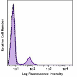
Each lot of this antibody is quality control tested by immunofluorescent staining with flow cytometric analysis. For flow cytometric staining, the suggested use of this reagent is ≤0.25 µg per million cells in 100 µl volume. It is recommended that the reagent be titrated for optimal performance for each application.

Excitation Laser
Blue Laser (488 nm)
Application Notes

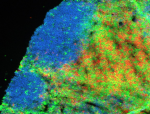
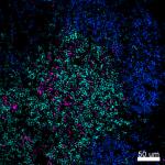
Additional reported applications (for the relevant formats) include: blocking of CD4+ T cell activation1,4,11, thymocyte costimulation3, in vitro and in vivo depletion2,5-8, blocking of egg-sperm cell adhesion1,4, immunohistochemical staining of acetone-fixed frozen sections9,10, immunoprecipitation1,2, and spatial biology (IBEX)12,13. The GK1.5 antibody is able to block CD4 mediated cell adhesion and T cell activation. Binding of GK1.5 antibody to CD4 T cells can be blocked by RM4-5 antibody, but not RM4-4 antibody. For in vivo studies or highly sensitive assays, we recommend Ultra-LEAF™ purified antibody (Cat. No. 100442) with a lower endotoxin limit than standard LEAF™ purified antibodies (Endotoxin < 0.01 EU/µg).

Application References
  1. Dialynas DP, et al. 1983. J. Immunol. 131:2445. (Block, IP)
  2. Dialynas DP, et al. 1983. Immunol. Rev. 74:29. (IP, Deplete)
  3. Wu L, et al. 1991. J. Exp. Med. 174:1617. (Costim)
  4. Godfrey DI, et al. 1994. J. Immunol. 152:4783. (Block)
  5. Gavett SH, et al. 1994. Am. J. Respir. Cell. Mol. Biol. 10:587. (Deplete)
  6. Schuyler M, et al. 1994. Am. J. Respir. Crit. Care Med. 149:1286. (Deplete)
  7. Ghobrial RR, et al. 1989. Clin. Immunol. Immunopathol. 52:486. (Deplete)
  8. Israelski DM, et al. 1989. J. Immunol. 142:954. (Deplete)
  9. Zheng B, et al. 1996. J. Exp. Med. 184:1083. (IHC)
  10. Frei K, et al. 1997. J. Exp. Med. 185:2177. (IHC)
  11. Felix NJ, et al. 2007. Nat. Immunol. 8:388. (Block)
  12. Radtke AJ, et al. 2020. Proc Natl Acad Sci U S A. 117:33455-65. (SB) PubMed
  13. Radtke AJ, et al. 2022. Nat Protoc. 17:378-401. (SB) PubMed
Product Citations
  1. Djokić V, et al. 2019. Front Immunol. 9:2891. PubMed
  2. Katsumura KR, et al. 2018. Proc Natl Acad Sci U S A. 115:E10109. PubMed
  3. Valanparambil RM, et al. 2017. PLoS Pathog. 13:e1006647. PubMed
  4. Xue G, et al. 2021. Cancer Cell. 39:1610. PubMed
  5. Mills EL, et al. 2022. Cell Metab. 34:140. PubMed
  6. Li X, et al. 2022. J Clin Invest. 132: . PubMed
  7. Wu R, et al. 2022. J Dermatol. 49:432. PubMed
  8. Mi Z, et al. 2021. Vaccines (Basel). 10: . PubMed
  9. Corral D, et al. 2022. Cell Rep. 39:110715. PubMed
  10. Lu R, et al. 2022. Scand J Immunol. 96:e13177. PubMed
  11. Shen Z, et al. 2022. EBioMedicine. 85:104285. PubMed
  12. Balood M, et al. 2022. Nature. 611:405. PubMed
  13. Zhu M, et al. 2022. Front Immunol. 13:1019248. PubMed
  14. Zhang Z, et al. 2022. Integr Cancer Ther. 21:15347354221138534. PubMed
  15. Wei M, et al. 2022. Front Immunol. 13:1017574. PubMed
  16. Wang H, et al. 2022. J Nanobiotechnology. 20:513. PubMed
  17. Chen C, et al. 2022. J Neuroinflammation. 19:315. PubMed
  18. Qin S, et al. 2023. Acta Pharm Sin B. :. PubMed
  19. Merk VM, et al. 2023. Genes Immun. 24:52. PubMed
  20. Meng S, et al. 2023. Stem Cell Investig. 10:2. PubMed
  21. Sun Y, et al. 2023. Cell Mol Life Sci. 80:63. PubMed
  22. Hajjar S, et al. 2022. Cell Death Differ. 29:585. PubMed
  23. Sylvester MA, et al. 2022. Am J Physiol Renal Physiol. 322:F245. PubMed
  24. Zander R, et al. 2022. Immunity. 55:475. PubMed
  25. Qiu Z, et al. 2022. J Exp Med. 219:. PubMed
  26. Lin J, et al. 2022. Adv Sci (Weinh). 9:e2202633. PubMed
  27. Hamdi L, et al. 2022. Ann Clin Transl Neurol. 9:1792. PubMed
  28. Deák P, et al. 2022. Cell Rep. 41:111563. PubMed
  29. Zhu C, et al. 2022. Chemotherapy. 67:211. PubMed
  30. Pan Y, et al. 2023. Adv Sci (Weinh). 10:e2206792. PubMed
  31. Wilson NG, et al. 2023. iScience. 26:105991. PubMed
  32. Zhuang WR, et al. 2023. Nat Commun. 14:1675. PubMed
  33. Mackin S, et al. 2023. Nat Microbiol. 8:569. PubMed
  34. Shi Y, et al. 2023. Nat Commun. 14:1884. PubMed
  35. Liu H, et al. 2023. Breast Cancer Res. 25:43. PubMed
  36. Fu S, et al. 2023. Nat Commun. 14:2248. PubMed
  37. Fiedler T, et al. 2023. J Neuroinflammation. 20:100. PubMed
  38. Yu L, et al. 2023. Front Oncol. 13:1174713. PubMed
  39. Wang Y, et al. 2023. iScience. 26:106630. PubMed
  40. Vereertbrugghen A, et al. 2023. J Neuroinflammation. 20:120. PubMed
  41. Peymanfar Y, et al. 2023. Int J Mol Sci. 24:. PubMed
  42. Wang X, et al. 2023. Nat Commun. 14:3440. PubMed
  43. Yao RQ, et al. 2023. Mil Med Res. 10:27. PubMed
  44. Iwanami N, et al. 2023. Nat Commun. 14:3645. PubMed
  45. Solvay M, et al. 2023. J Immunother Cancer. 11:. PubMed
  46. Wang Z, et al. 2023. Sci Adv. 9:eabo4100. PubMed
  47. Zelenka T, et al. 2022. Nat Commun. 13:6954. PubMed
  48. Zhao J, et al. 2021. Emerg Microbes Infect. 10:913. PubMed
  49. Liu J, et al. 2021. Front Immunol. 12:747730. PubMed
  50. Su Y, et al. 2022. J Hematol Oncol. 15:99. PubMed
  51. Li Z, et al. 2022. Nat Commun. 13:1845. PubMed
  52. Chang YS, et al. 2020. Int J Mol Sci. 21:00. PubMed
  53. Chuang H, et al. 2014. Nat Commun. 5:4602. PubMed
  54. Wang X, et al. 2019. Cell Res. 29:787. PubMed
  55. Alissafi T, et al. 2018. J Clin Invest. 128:3840. PubMed
  56. Yang Z, et al. 2022. J Immunother Cancer. 10:. PubMed
  57. Li Q, et al. 2022. Cell Rep. 40:111308. PubMed
  58. Schönberger K, et al. 2022. Cell Stem Cell. 29:131. PubMed
  59. He R, et al. 2021. J Immunol. 206:2353. PubMed
  60. Ma J, et al. 2020. Adv Sci (Weinh). 7:2000609. PubMed
  61. Fan L, et al. 2021. Cancers (Basel). 13:. PubMed
  62. Rosenkranz SC, et al. 2021. eLife. 10:00. PubMed
  63. Furuta Y, et al. 2017. PLoS One. 12(2):e0172509. PubMed
  64. Wu N, et al. 2020. Cell Rep. 30:1129. PubMed
  65. Saleh MM, et al. 2019. Cell Host Microbe. 25:756. PubMed
  66. Toomer G, et al. 2022. Viruses. 14:. PubMed
  67. Luo R, et al. 2021. Theranostics. 11:117. PubMed
  68. Xu ZH, et al. 2021. Mediators Inflamm. 2021:8856326. PubMed
  69. Liang S, et al. 2022. Acta Pharm Sin B. 12:2494. PubMed
  70. Jiang W, et al. 2022. Front Immunol. 13:878832. PubMed
  71. Yang F, et al. 2021. Front Microbiol. 11:512581. PubMed
  72. Sega E, et al. 2008. Int J Radiat Oncol Biol Phys. 71:559. PubMed
  73. Lua Y, et al. 2012. Mol Immunol. 52:229. PubMed
  74. Zasłona Z, et al. 2014. J Immunol. 193:4245. PubMed
  75. Aguilera T, et al. 2016. Nat Commun. 7:13898. PubMed
  76. Periasamy S, et al. 2017. Nat Commun. 8:15564. PubMed
  77. Pobezinskaya EL et al. 2019. Front Immunol. 1.079861111 . PubMed
  78. Choi H et al. 2019. Cell Rep. 27(3):806-819 . PubMed
  79. Liang Q, et al. 2022. iScience. 25:104043. PubMed
  80. Mirshahi F, et al. 2022. Cell Rep. 38:110454. PubMed
  81. Bangs DJ, et al. 2022. Cell Rep. 38:110266. PubMed
  82. Jazowiecka-Rakus J, et al. 2021. Cancers (Basel). 13:. PubMed
  83. Chen J, et al. 2022. J Nanobiotechnology. 20:283. PubMed
  84. Wu M, et al. 2021. Nat Commun. 12:3500. PubMed
  85. Fu Y, et al. 2020. Mol Ther. 1214:28. PubMed
  86. Altshuler A, et al. 2021. Cell Stem Cell. 28(7):1248-1261.e8. PubMed
  87. Tang Y, et al. 2020. Front Immunol. 11:585918. PubMed
  88. Qi X, et al. 2019. Nat Commun. 10:2141. PubMed
  89. Shi S, et al. 2020. Clin Cancer Res. 26:5990. PubMed
  90. Wang Y, et al. 2022. Acta Pharm Sin B. 12:262. PubMed
  91. Zhou HF, et al. 2022. Front Pharmacol. 13:778755. PubMed
  92. Liang Z, et al. 2021. J Cell Physiol. 236:4725. PubMed
  93. Mardomi A, et al. 2021. J Cell Physiol. 236:6328. PubMed
  94. Xu F, et al. 2022. Cell Death Discov. 8:142. PubMed
  95. He C, et al. 2020. Clin Transl Med. 10:e39. PubMed
  96. Molina-Sanchez P, et al. 2020. Gastroenterology. 2203:159. PubMed
  97. Brooks JF, et al. 2020. Cytotherapy. 22:436. PubMed
  98. Aqel SI, et al. 2021. JCI Insight. 6:e142376. PubMed
  99. Hu D, et al. 2021. J Cell Mol Med. 25:2900. PubMed
  100. D Kim, S Lee, H Jun 2016. Immunol Cell Biol. 10.1038/icb.2016.98. PubMed
  101. Wu J, et al. 2020. Sci Adv. 6:eaba3458. PubMed
  102. Guan H, et al. 2012. PLoS One. 7:e35650. PubMed
  103. Thomas S, et al. 2019. J Immunother Cancer. 7:214. PubMed
  104. Tritama E, et al. 2018. Hum Vaccin Immunother. 14:1524. PubMed
  105. Mauduit O, et al. 2022. Front Immunol. 13:1011125. PubMed
  106. Liu Y, et al. 2022. Nat Commun. 13:2665. PubMed
  107. Anastasiou M, et al. 2021. JCI Insight. 6:. PubMed
  108. Li H, et al. 2021. Nat Commun. 12:2773. PubMed
  109. Jiang S, et al. 2020. Scand J Immunol. e12867:91. PubMed
  110. Mitchell JE, et al. 2021. Cell Reports. 35(2):108966. PubMed
  111. White C, et al. 2014. J Immunol. 193:5933. PubMed
  112. Sarapulov AV, et al. 2020. Front Immunol. 11:599. PubMed
  113. Zongyi Y, et al. 2017. PLoS One. 10.1371/journal.pone.0189617. PubMed
  114. Ren HZ, et al. 2022. J Clin Transl Hepatol. 10:1138. PubMed
  115. Edwards CJ, et al. 2021. Br J Cancer. . PubMed
  116. Jing L, et al. 2022. Front Immunol. 13:864995. PubMed
  117. Feng H, et al. 2020. J Dermatol. 47:147. PubMed
  118. Ma X, et al. 2021. Cell Metabolism. 33(5):1001-1012.e5. PubMed
  119. Cheng K, et al. 2021. Nat Commun. 12:2041. PubMed
  120. Pedraza–Zamora CP, et al. 2017. Parasite Immunol. 39:4. PubMed
  121. Raineri D, et al. 2021. Biomedicines. 10:. PubMed
  122. He X, et al. 2021. Small. 17:e2007165. PubMed
  123. Katsinas N, et al. 2022. J Clin Med. 11:. PubMed
  124. He X, et al. 2021. Adv Sci (Weinh). 8:e2103023. PubMed
  125. Chen L, et al. 2021. Cell Death Differ. 28:1880. PubMed
  126. Wang Z, et al. 2020. J Clin Invest. 130:3717. PubMed
  127. Zhang W, et al. 2019. Oncol Lett. 17:815. PubMed
  128. Cheng B, et al. 2022. Cancer Commun (Lond). 42:17. PubMed
  129. Wang Z, et al. 2022. Nat Biomed Eng. 6:791. PubMed
  130. Guo P, et al. 2021. J Immunol. 207:408. PubMed
  131. Deng Z, et al. 2020. World J Gastrointest Oncol. 1091:12. PubMed
  132. Vigeland C, et al. 2016. PLoS One. 11: 0163288. PubMed
  133. Lu X, et al. 2016. Nat Commun. 7: 12719. PubMed
  134. Cervera–Carrascon V, et al. 2020. Oncoimmunology. 9:1761229. PubMed
  135. Paul S, et al. 2018. Appl Microbiol Biotechnol. 102:8895. PubMed
  136. Dmitrieva‐Posocco O et al. 2019. Immunity. 50(1):166-180 . PubMed
  137. Kim JH, et al. 2019. Mol Med. 25:33. PubMed
  138. Niknam S, et al. 2018. Clin Cancer Res. 24:5735. PubMed
  139. Habib S, et al. 2018. Infect Immun. 86:e00019. PubMed
  140. Zhao X, et al. 2022. Nat Protoc. 17:2240. PubMed
  141. Zhou H, et al. 2022. Oncoimmunology. 11:2057892. PubMed
  142. He X, et al. 2022. Cancer Immunol Res. 10:314. PubMed
  143. Hum NR, et al. 2022. Front Oncol. 12:928474. PubMed
  144. Zeng Q, et al. 2022. Front Immunol. 13:740805. PubMed
  145. Zuo S, et al. 2021. J Immunother Cancer. 9:. PubMed
  146. Zhang Y, et al. 2021. J Immunol Res. 2021:5599439. PubMed
  147. Volberding PJ, et al. 2021. Cell Reports. 35(8):109160. PubMed
  148. Knocke S, et al. 2016. Cell Rep. 17:2234-2246. PubMed
  149. Ahmari N, et al. 2016. Physiol Genomics. 48: 526 - 536. PubMed
  150. Ito C, et al. 2015. PLoS One. 10: e0140808. PubMed
  151. Zhu Y, et al. 2021. EMBO J. 40:e105320. PubMed
  152. Wang Y, et al. 2021. Front Microbiol. 12:701566. PubMed
  153. Gebre MS, et al. 2022. NPJ Vaccines. 7:88. PubMed
  154. Yang B, et al. 2022. Bioengineered. 13:2685. PubMed
  155. Faust HJ, et al. 2020. J Clin Invest. 130:5493. PubMed
  156. Kruta M, et al. 2021. Cell Stem Cell. :. PubMed
  157. Haas MS, et al. 2020. Mol Cancer Res. 19:717. PubMed
  158. Uddback I, et al. 2016. Sci Rep. 6:20137. PubMed
  159. Toomer K, et al. 2016. J Immunol. 196: 3665 - 3676. PubMed
  160. Zhang H, et al. 2019. Mol Cell. 76:110. PubMed
  161. Zhou J, et al. 2019. Nat Commun. 10:2427. PubMed
  162. Sun H, et al. 2022. Front Immunol. 13:818017. PubMed
  163. Frost JN, et al. 2021. Med (N Y). 2:164. PubMed
  164. Ding P, et al. 2022. Bone Res. 10:42. PubMed
  165. Lang V, et al. 2021. Elife. 10:. PubMed
  166. Peng L, et al. 2022. Cell Rep Med. 3:100634. PubMed
  167. Huang L, et al. 2021. Breast Cancer Res Treat. Online ahead of print. PubMed
  168. Lu J, et al. 2021. Cell Death Discov. 7:165. PubMed
  169. Han F, et al. 2020. Invest Ophthalmol Vis Sci. 61:24. PubMed
  170. Yan J, et al. 2020. Cell Rep. 107820:31. PubMed
  171. Shannon JP, et al. 2021. Immunity. 54(2):276-290.e5. PubMed
  172. Mencl S, et al. 2014. J Neuroimmunol. 274:125. PubMed
  173. Subramanian K, et al. 2019. Nat Microbiol. 4:62. PubMed
  174. Yang BH, et al. 2020. Cell Reports. 27(12):3629-3645.e6.. PubMed
  175. Zeng Q, et al. 2022. iScience. 25:105151. PubMed
  176. Guo S, et al. 2021. Sci Rep. 11:23745. PubMed
  177. Ma K, et al. 2022. iScience. 25:104347. PubMed
  178. Li J, et al. 2022. Nat Commun. 13:4032. PubMed
  179. Gao X, et al. 2022. Front Immunol. 13:799331. PubMed
  180. Timilshina M, et al. 2017. PLoS One. 10.1371/journal.pone.0168942. PubMed
  181. Wang X, et al. 2020. Nat Commun. 4.505555556. PubMed
  182. Wei Z, et al. 2021. Nat Commun. 0.805555556. PubMed
  183. Zhang Q, et al. 2021. Front Cell Dev Biol. 9:655552. PubMed
  184. Kmieciak M, et al. 2011. J Vis Exp. 47: 2381. PubMed
  185. Li Y, Kaneda T 2016. Sci Rep. 6: 25077. PubMed
  186. Tao Z, et al. 2019. Clin Cancer Res. 25:1113. PubMed
  187. Reynolds CJ, et al. 2018. Sci Rep. 8:672. PubMed
  188. Li Z, et al. 2022. Nat Commun. 13:6321. PubMed
  189. He C, et al. 2022. Nat Commun. 13:5459. PubMed
  190. Takaku S, et al. 2022. Evid Based Complement Alternat Med. 2022:6122955. PubMed
  191. Zhang S, et al. 2022. Nat Commun. 13:4744. PubMed
  192. Yuan M, et al. 2022. Oxid Med Cell Longev. 2022:5479491. PubMed
  193. Li J, et al. 2020. Development. :. PubMed
  194. Shibuya M, et al. 2021. iScience. 24:103131. PubMed
  195. Nasarre P, et al. 2021. Cancers (Basel). 13: . PubMed
  196. Wang Y, et al. 2020. Virol Sin. 36:122. PubMed
  197. Liu Y, et al. 2020. Oncol Lett. 2369:20. PubMed
  198. Martomo SA, et al. 2021. Mol Cancer Ther. 1.074305556. PubMed
  199. Zan H, et al. 2017. Nat Commun. 8:14244. PubMed
  200. Kamata T, et al. 2020. Cell Reports. 31(12):107802. PubMed
  201. Ouyang S, et al. 2019. J Immunol. 202:1441. PubMed
  202. Nechama M, et al. 2018. Nat Commun. 9:1603. PubMed
  203. Xia Y, et al. 2018. Cell. 175:1059. PubMed
  204. Karanika S, et al. 2022. Front Immunol. 13:972266. PubMed
  205. Zhang Q, et al. 2022. Commun Biol. 5:544. PubMed
  206. Jazowiecka-Rakus J, et al. 2020. Mol Ther Oncolytics. 18:335. PubMed
  207. Soria-Castro R, et al. 2020. Sci Rep. 10:17802. PubMed
  208. Chen H, et al. 2022. Front Pharmacol. 13:827520. PubMed
  209. Ligon MM, et al. 2020. Mucosal Immunol. 1.172222222. PubMed
  210. Da Mesquita S, et al. 2021. Science Advances. 7(21):. PubMed
  211. Wedekind MF, et al. 2021. iScience. 24(7):102759. PubMed
  212. Liu YG, et al. 2019. FASEB J. 33:5018. PubMed
  213. Yao C, et al. 2018. Autophagy. 1.854861111. PubMed
  214. Ji L et al. 2019. Immunity. 51(2):272-284 . PubMed
  215. Habtetsion T et al. 2018. Cell metabolism. 28(2):228-242 . PubMed
  216. Coley WD et al. 2018. F1000Research. 2018 Mar 14 [revised 2018 Jan 1] 0.5125. PubMed
  217. Pan L, et al. 2022. Front Immunol. 13:837497. PubMed
  218. Zheng Y, et al. 2022. J Immunol. 208:501. PubMed
  219. He X, et al. 2021. J Immunother Cancer. 9:. PubMed
  220. Zhou H, et al. 2021. Cell Death Discov. 7:332. PubMed
  221. Liu Y, et al. 2020. Cell Death Dis. 11:1062. PubMed
  222. Si J, et al. 2020. Cancer Cell. 38(4):551-566.e11. PubMed
  223. Khosravi N, et al. 2018. Cancer Immunol Res. 0.797222222. PubMed
  224. Kos S, et al. 2019. PLoS One. 14:e0217762. PubMed
  225. Golan K et al. 2018. Cell stem cell. 23(4):572-585 . PubMed
  226. Sears SM, et al. 2022. Kidney360. 3:818. PubMed
  227. Sun F, et al. 2022. Cell Death Dis. 13:181. PubMed
  228. Zhong C, et al. 2021. J Virol. 95:e0092521. PubMed
  229. Magee JA, et al. 2021. Stem Cell Reports. 16:20. PubMed
  230. Xu Y, et al. 2021. iScience. 24:103445. PubMed
  231. Zhou S, et al. 2021. Adv Funct Mater. 31:. PubMed
  232. Smith KJ, et al. 2022. PLoS Biol. 20:e3001554. PubMed
  233. Blaszczak AM, et al. 2020. Immunometabolism. 2:00. PubMed
  234. Yang C, et al. 2020. Cell Host Microbe. 467:27. PubMed
  235. Bryan Hart, Sunhee Lee 2016. PLoS Negl Trop Dis. 10:e0005229. PubMed
  236. Sivapatham S, et al. 2019. Front Immunol. 1.865277778. PubMed
  237. Zhao Y, et al. 2020. Front Immunol. 2.572222222. PubMed
  238. Blaszczak AM, et al. 2019. J Immunol. 202:2451. PubMed
  239. Liang Z, et al. 2017. Autophagy. 14:505. PubMed
  240. Nakao R, et al. 2022. NPJ Vaccines. 7:153. PubMed
  241. Wang Y, et al. 2022. Nat Commun. 13:1454. PubMed
  242. Barsoumian HB, et al. 2022. Cancers (Basel). 14:. PubMed
  243. Chen YL, et al. 2022. Front Neurosci. 16:876582. PubMed
  244. Li Z, et al. 2022. Evid Based Complement Alternat Med. 2022:7421265. PubMed
  245. Yue T, et al. 2022. Cell Death Differ. 29:218. PubMed
  246. Aguilera TA, et al. 2020. Clin Cancer Res. 26:2972. PubMed
  247. Simoni L, et al. 2020. Cell Rep. 33:108330. PubMed
  248. Hart B, et al. 2015. PLoS Negl Trop Dis. 9: 0004046. PubMed
  249. Ramanan D, et al. 2020. Cell. 181(6):1276-1290. PubMed
  250. Hidalgo San Jose L, et al. 2020. Cell Rep. 30:69. PubMed
  251. Bartley A, et al. 2018. Front Physiol. 9:1593. PubMed
  252. Timilshina M, et al. 2020. Cell Reports. 27(10):2948-2961.e7.. PubMed
  253. Hsu HP, et al. 2021. J Biol Chem. 296:100419. PubMed
  254. Zhang M, et al. 2022. iScience. 25:104490. PubMed
  255. Chen Y, et al. 2021. Braz J Med Biol Res. 54:e9570. PubMed
  256. Rodrigues KA, et al. 2021. Sci Adv. 7:eabj6538. PubMed
  257. Kathania M, et al. 2022. Cell Rep. 40:111345. PubMed
  258. Gawish R, et al. 2022. Elife. 11:. PubMed
  259. Li J, et al. 2022. Nat Commun. 13:1481. PubMed
  260. Gardenier J, et al. 2017. Nat Commun. 8:14345. PubMed
  261. Jaeger N, et al. 2020. Cell Rep. 33:108331. PubMed
  262. Zeng A, et al. 2020. Oncol Rep. . PubMed
  263. Angela M, et al. 2016. Nat Commun. 7:13683. PubMed
  264. Hong Y, et al. 2019. J Extracell Vesicles. 8:1670893. PubMed
  265. Zhou T, et al. 2018. Mol Med Rep. 17:4883. PubMed
  266. Li CY, et al. 2022. Int J Mol Sci. 23:. PubMed
  267. Liang Z, et al. 2022. iScience. 25:105233. PubMed
  268. Lestari SR, et al. 2020. J Ayurveda Integr Med. 11:414. PubMed
  269. Jiang W, et al. 2021. Cell Rep. 37:110112. PubMed
  270. Chang MH, et al. 2021. Cell Rep. 37:109902. PubMed
  271. Ding X, et al. 2021. Front Immunol. 12:667136. PubMed
  272. Guo S, et al. 2021. Frontiers in Immunology. 11:614667. PubMed
  273. Cortez MA, et al. 2020. Nat Commun. 3.819444444. PubMed
RRID
AB_312690 (BioLegend Cat. No. 100405)
AB_312691 (BioLegend Cat. No. 100406)

Antigen Details

Structure
Ig superfamily, 55 kD
Distribution

Majority of thymocytes, T cell subset

Function
TCR co-receptor, T cell activation
Ligand/Receptor
MHC class II molecule
Cell Type
Dendritic cells, T cells, Thymocytes, Tregs
Biology Area
Immunology
Molecular Family
CD Molecules
Antigen References

1. Barclay A, et al. 1997. The Leukocyte Antigen FactsBook Academic Press.
2. Bierer BE, et al. 1989. Annu. Rev. Immunol. 7:579.
3. Janeway CA. 1992. Annu. Rev. Immunol. 10:645.

Gene ID
12504 View all products for this Gene ID
UniProt
View information about CD4 on UniProt.org

Related FAQs

I am unable to see expression of T cell markers such as CD3 and CD4 post activation.
TCR-CD3 complexes on the T-lymphocyte surface are rapidly downregulated upon activation with peptide-MHC complex, superantigen or cross-linking with anti-TCR or anti-CD3 antibodies. PMA/Ionomycin treatment has been shown to downregulate surface CD4 expression. Receptor downregulation is a common biological phenomenon and so make sure that your stimulation treatment is not causing it in your sample type.

Other Formats

View All CD4 Reagents Request Custom Conjugation
Description Clone Applications
APC anti-mouse CD4 GK1.5 FC
Biotin anti-mouse CD4 GK1.5 FC,IHC-F,ICC
FITC anti-mouse CD4 GK1.5 FC,IHC-F,ICC
PE anti-mouse CD4 GK1.5 FC
PE/Cyanine5 anti-mouse CD4 GK1.5 FC
Purified anti-mouse CD4 GK1.5 FC,IHC-F,ICC,IP,Costim,Block,Depletion
PE/Cyanine7 anti-mouse CD4 GK1.5 FC
APC/Cyanine7 anti-mouse CD4 GK1.5 FC
Alexa Fluor® 647 anti-mouse CD4 GK1.5 FC,IHC-F,ICC
Alexa Fluor® 488 anti-mouse CD4 GK1.5 FC,IHC-F,ICC
Pacific Blue™ anti-mouse CD4 GK1.5 FC
Alexa Fluor® 700 anti-mouse CD4 GK1.5 FC
PerCP anti-mouse CD4 GK1.5 FC
PerCP/Cyanine5.5 anti-mouse CD4 GK1.5 FC
Brilliant Violet 421™ anti-mouse CD4 GK1.5 FC,ICC,IHC-F
Ultra-LEAF™ Purified anti-mouse CD4 GK1.5 FC,Block,Costim,Depletion,IHC,IP
Alexa Fluor® 594 anti-mouse CD4 GK1.5 IHC-F,FC,ICC,SB
Brilliant Violet 711™ anti-mouse CD4 GK1.5 FC
Brilliant Violet 510™ anti-mouse CD4 GK1.5 FC,IHC-F,ICC
Brilliant Violet 605™ anti-mouse CD4 GK1.5 FC
Brilliant Violet 785™ anti-mouse CD4 GK1.5 FC
PE/Dazzle™ 594 anti-mouse CD4 GK1.5 FC
APC/Fire™ 750 anti-mouse CD4 GK1.5 FC
GoInVivo™ Purified anti-mouse CD4 GK1.5 FC
Brilliant Violet 750™ anti-mouse CD4 GK1.5 FC
Brilliant Violet 650™ anti-mouse CD4 GK1.5 FC
Spark Blue™ 550 anti-mouse CD4 GK1.5 FC
Spark NIR™ 685 anti-mouse CD4 GK1.5 FC
KIRAVIA Blue 520™ anti-mouse CD4 GK1.5 FC
PE/Fire™ 640 anti-mouse CD4 GK1.5 FC
APC/Fire™ 810 anti-mouse CD4 GK1.5 FC
PE/Fire™ 700 anti-mouse CD4 GK1.5 FC
Spark Violet™ 538 anti-mouse CD4 GK1.5 FC
Spark YG™ 593 anti-mouse CD4 GK1.5 FC
Spark Blue™ 574 anti-mouse CD4 Antibody GK1.5 FC
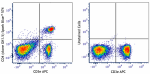
Spark UV™ 387 anti-mouse CD4 GK1.5 FC
Spark Blue™ 515 anti-mouse CD4 GK1.5 FC
Spark PLUS UV395™ anti-mouse CD4 GK1.5 FC
Spark Red™ 718 anti-mouse CD4 (Flexi-Fluor™) GK1.5 FC
PerCP/Fire™ 806 anti-mouse CD4 Antibody GK1.5 FC
PerCP/Fire™ 780 anti-mouse CD4 Antibody GK1.5 FC
Go To Top Version: 1    Revision Date: 11/30/2012

For Research Use Only. Not for diagnostic or therapeutic use.


This product is supplied subject to the terms and conditions, including the limited license, located at www.biolegend.com/terms) ("Terms") and may be used only as provided in the Terms. Without limiting the foregoing, BioLegend products may not be used for any Commercial Purpose as defined in the Terms, resold in any form, used in manufacturing, or reverse engineered, sequenced, or otherwise studied or used to learn its design or composition without express written approval of BioLegend. Regardless of the information given in this document, user is solely responsible for determining any license requirements necessary for user’s intended use and assumes all risk and liability arising from use of the product. BioLegend is not responsible for patent infringement or any other risks or liabilities whatsoever resulting from the use of its products.


BioLegend, the BioLegend logo, and all other trademarks are property of BioLegend, Inc. or their respective owners, and all rights are reserved.


8999 BioLegend Way, San Diego, CA 92121 www.biolegend.com
Toll-Free Phone: 1-877-Bio-Legend (246-5343) Phone: (858) 768-5800 Fax: (877) 455-9587

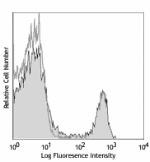
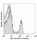
This data display is provided for general comparisons between formats.
Your actual data may vary due to variations in samples, target cells, instruments and their settings, staining conditions, and other factors.
If you need assistance with selecting the best format contact our expert technical support team.

ProductsHere

Login / Register
Remember me
Forgot your password? Reset password?
Create an Account