 Login / Register


 United States ($) USD
 Austria (€) EUR
 Belgium (€) EUR
 Finland (€) EUR
 France (€) EUR
 Germany (€) EUR
 Ireland (€) EUR
 Japan (¥) JPY
 Luxembourg (€) EUR
 Netherlands (€) EUR
 Switzerland (CHF) CHF
 United Kingdom (£) GBP

- Clone
- CD28.2 (See other available formats)
- Regulatory Status
- RUO
- Workshop
- V-CD28.05
- Other Names
- T44, Tp44
- Isotype
- Mouse IgG1, κ
- Ave. Rating
- Submit a Review
- Product Citations
- publications
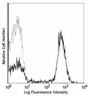
-

Human peripheral blood lymphocytes stained with CD28.2 PE
| Cat # | Size | Price | Quantity Check Availability | Save | ||
|---|---|---|---|---|---|---|
| 302901 | 25 µg | 20€ | ||||
| 302902 | 100 µg | 40€ | ||||
CD28 is a 44 kD disulfide-linked homodimeric type I glycoprotein. It is a member of the immunoglobulin superfamily and is also known as T44 or Tp44. CD28 is expressed on most T lineage cells, NK cell subsets, and plasma cells. CD28 binds both CD80 and CD86 using a highly conserved motif MYPPY in the CDR3-like loop. CD28 is considered a major co-stimulatory molecule, inducing T lymphocyte activation and IL-2 synthesis, and preventing cell death. In vitro studies indicate that ligation of CD28 on T cells by CD80 and CD86 on antigen presenting cells provides a costimulatory signal required for T cell activation and proliferation.
Product DetailsProduct Details
- Verified Reactivity
- Human, Cynomolgus, Rhesus
- Reported Reactivity
- Baboon, Capuchin Monkey, Chimpanzee, Pigtailed Macaque, Sooty Mangabey, Squirrel Monkey
- Antibody Type
- Monoclonal
- Host Species
- Mouse
- Formulation
- Phosphate-buffered solution, pH 7.2, containing 0.09% sodium azide.
- Preparation
- The antibody was purified by affinity chromatography.
- Concentration
- 0.5 mg/ml
- Storage & Handling
- The antibody solution should be stored undiluted between 2°C and 8°C.
- Application
-
FC - Quality tested
IHC-F, Costim, FA - Reported in the literature, not verified in house - Recommended Usage
-
Each lot of this antibody is quality control tested by immunofluorescent staining with flow cytometric analysis. For flow cytometric staining, the suggested use of this reagent is ≤ 0.5 µg per 106 cells in 100 µl volume or 100 µl whole blood. It is recommended that the reagent be titrated for optimal performance for each application.
- Application Notes
-
The Ultra-LEAF™ Purified antibody (Endotoxin < 0.01 EU/µg, Azide-Free, 0.2 µm filtered) is recommended for highly sensitive assays.
-
Application References
(PubMed link indicates BioLegend citation) -
- Schlossman S, et al. Eds. 1995. Leucocyte Typing V. Oxford University Press. New York.
- Nunes J, et al. 1993. Biochem. J. 293:835.
- Calea-Lauri J, et al. 1999. J. Immunol. 163:62.
- Tazi A, et al. 1999. J. Immunol. 163:3511. (IHC)
- Marti F, et al. 2001. J. Immunol. 166:197. (Costim)
- Jeong SH, et al. 2004. J. Virol. 78:6995. (Costim)
- Rivollier A, et al. 2004. Blood 104:4029. (Costim)
- Scharschmidt E, et al. 2004. Mol. Cell Biol. 24:3860. (Costim)
- Sheng W, et al. 2007.Elsevier 580:6819. PubMed
- Mitsuhashi M. 2007. Clin Chem.53:148. PubMed
- Ye Z, et al. 2008. Infect. Immun. 76:2541. PubMed
- Magatti M, et al. 2008. Stem Cells 26:182. (FA) PubMed
- Yoshino N, et al. 2008. Exp. Anim. (Tokyo) 49:97. (FC)
- Berg M, et al. 2008. J Leukoc Biol. 83:853. (IP) PubMed
- Rout N, et al. 2010. PLoS One 5:e9787. (FC)
- Leonard JA, et al. 2011. J. Virol. 85:6867. PubMed
- Nomura T, et al. 2012. J. Virol. 86:6481. PubMed
- Product Citations
-
- RRID
-
AB_314303 (BioLegend Cat. No. 302901)
AB_314304 (BioLegend Cat. No. 302902)
Antigen Details
- Structure
- Ig superfamily, type I transmembrane glycoprotein, homodimer, 44 kD
- Distribution
-
Mature T cells, thymocytes, NK cell subsets, plasma cells, EBV-positive B cells
- Function
- T cell costimulation
- Ligand/Receptor
- CD80, CD86
- Cell Type
- B cells, NK cells, Plasma cells, T cells, Thymocytes, Tregs
- Biology Area
- Costimulatory Molecules, Immunology
- Molecular Family
- CD Molecules
- Antigen References
-
1. Schlossman S, et al. Eds. 1995. Leucocyte Typing V. Oxford University Press. New York.
2. June CH, et al. 1994. Immunol. Today 15:321.
3. Linskey PS, et al. 1993. Annu. Rev. Immunol. 11:191. - Gene ID
- 940 View all products for this Gene ID
- Specificity (DOES NOT SHOW ON TDS):
- CD28
- Specificity Alt (DOES NOT SHOW ON TDS):
- CD28
- App Abbreviation (DOES NOT SHOW ON TDS):
- FC,IHC-F,Costim,FA
- UniProt
- View information about CD28 on UniProt.org


Related FAQs
Other Formats
View All CD28 Reagents Request Custom ConjugationCustomers Also Purchased
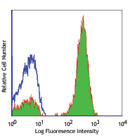
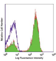
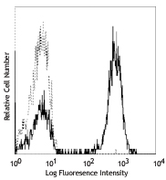
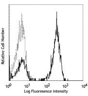
Compare Data Across All Formats
This data display is provided for general comparisons between formats.
Your actual data may vary due to variations in samples, target cells, instruments and their settings, staining conditions, and other factors.
If you need assistance with selecting the best format contact our expert technical support team.
-
APC anti-human CD28
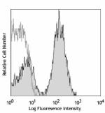
Human peripheral blood lymphocytes stained with APC CD28.2 -
Biotin anti-human CD28
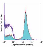
Human peripheral blood lymphocytes stained with biotinylated... -
FITC anti-human CD28
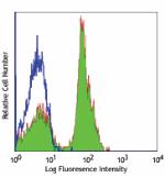
Human peripheral blood lymphocytes stained with CD28.2 FITC -
PE anti-human CD28
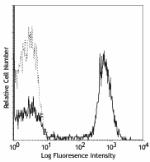
Human peripheral blood lymphocytes stained with CD28.2 PE -
PE/Cyanine5 anti-human CD28
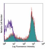
Human peripheral blood lymphocytes stained with CD28.2 PE/Cy... -
Purified anti-human CD28
Human peripheral blood lymphocytes stained with CD28.2 PE -
Alexa Fluor® 488 anti-human CD28
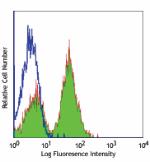
Human peripheral blood lymphocytes stained with CD28.2 Alexa... -
Alexa Fluor® 700 anti-human CD28
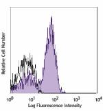
Human peripheral blood lymphocytes stained with CD28.2 Alexa... -
PerCP/Cyanine5.5 anti-human CD28
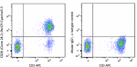
Human peripheral blood lymphocytes were stained with CD3 APC... -
Pacific Blue™ anti-human CD28
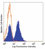
Human peripheral blood lymphocytes stained with CD28.2 Pacif... -
PE/Cyanine7 anti-human CD28
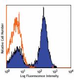
Human peripheral blood lymphocytes stained with CD28.2 PE/Cy... -
Ultra-LEAF™ Purified anti-human CD28
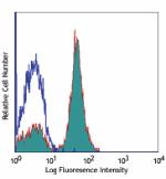
Human peripheral blood lymphocytes stained with LEAF™ purifi... 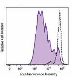
Human peripheral blood mononuclear cells were stained with C... 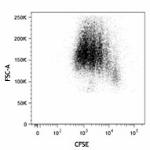
Human peripheral blood mononuclear cells were stained with C... -
Brilliant Violet 421™ anti-human CD28
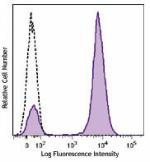
Human peripheral blood lymphocytes were stained with CD28 (c... -
Brilliant Violet 510™ anti-human CD28
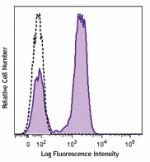
Human peripheral blood lymphocytes were stained with CD28 (c... -
Purified anti-human CD28 (Maxpar® Ready)
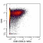
Human PBMCs stained with 154Sm-anti-CD45 (HI30) and 160Gd-an... -
PE/Dazzle™ 594 anti-human CD28
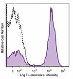
Human peripheral blood lymphocytes were stained with CD28 (c... -
Brilliant Violet 785™ anti-human CD28
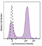
Human peripheral blood lymphocytes were stained with CD28 (c... -
Brilliant Violet 650™ anti-human CD28
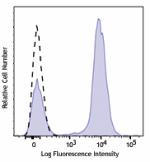
Human peripheral blood Lymphocytes were stained with CD28 (c... -
Brilliant Violet 711™ anti-human CD28
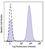
Human peripheral blood Lymphocytes were stained with CD28 (c... -
APC/Fire™ 750 anti-human CD28
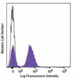
Human peripheral blood lymphocytes were stained with CD28 (c... -
Alexa Fluor® 647 anti-human CD28
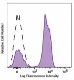
Human peripheral blood lymphocytes were stained with CD28 (c... -
TotalSeq™-A0386 anti-human CD28
-
TotalSeq™-B0386 anti-human CD28
-
TotalSeq™-C0386 anti-human CD28
-
Brilliant Violet 605™ anti-human CD28
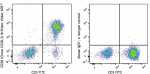
Human peripheral blood lymphocytes were stained with CD3 FIT... -
APC/Cyanine7 anti-human CD28
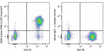
Human peripheral blood lymphocytes were stained with True-St... -
PE anti-human CD28

Typical results from human peripheral blood lymphocytes stai... -
APC anti-human CD28

Typical results from human peripheral blood lymphocytes stai... -
Brilliant Violet 750™ anti-human CD28
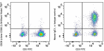
Human peripheral blood lymphocytes were stained with CD3 FIT... -
PE/Fire™ 810 anti-human CD28
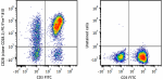
Human peripheral blood lymphocytes were stained with anti-hu... -
GMP PE anti-human CD28
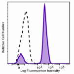
Typical results from human peripheral blood lymphocytes stai... -
TotalSeq™-D0386 anti-human CD28
-
Spark Violet™ 423 anti-human CD28
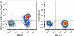
Human peripheral blood lymphocytes were stained with anti-hu... -
GMP Ultra-LEAF™ Purified anti-human CD28 SF
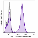
Human peripheral blood lymphocytes were stained with GMP Ult... 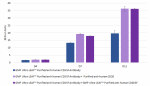
PBMC-derived T cells were activated in the presence of GMP U... -
GMP APC anti-human CD28
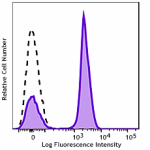
Typical results from human peripheral blood lymphocytes stai... -
PE/Cyanine7 anti-human CD28
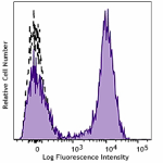
Typical results from human peripheral blood lymphocytes stai... -
Cell-Vive™ GMP Ultra-LEAF™ Biotin anti-human CD28 SF
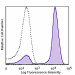
Human peripheral lymphocytes were stained with GMP Ultra-LEA... 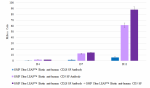
PBMC-derived T cells were activated in the presence of GMP U... -
PerCP/Cyanine5.5 anti-human CD28
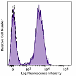
Typical results from human peripheral blood lymphocytes stai... -
Spark Blue™ 515 anti-human CD28
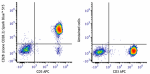
Human peripheral blood lymphocytes were stained with anti-hu... -
Spark Red™ 718 anti-human CD28
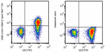
Human peripheral blood lymphocytes were stained with anti-hu... -
GMP PerCP/Cyanine5.5 anti-human CD28

Typical results from human peripheral blood lymphocytes stai... -
Pacific Blue™ anti-human CD28

Typical results from human peripheral blood lymphocytes stai... -
APC/Fire™ 810 anti-human CD28

Human peripheral blood lymphocytes were stained with anti-hu...


Follow Us