 Login / Register


 United States ($) USD
 Austria (€) EUR
 Belgium (€) EUR
 Finland (€) EUR
 France (€) EUR
 Germany (€) EUR
 Ireland (€) EUR
 Japan (¥) JPY
 Luxembourg (€) EUR
 Netherlands (€) EUR
 Switzerland (CHF) CHF
 United Kingdom (£) GBP

- Clone
- C068C2 (See other available formats)
- Regulatory Status
- RUO
- Other Names
- MMR (macrophage mannose receptor), MR (mannose receptor), MRC1
- Isotype
- Rat IgG2a, κ
- Ave. Rating
- Submit a Review
- Product Citations
- publications
-

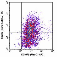
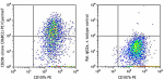
Thioglycollate-elicited BALB/c peritoneal macrophages were surface stained with CD107b (Mac-3) APC, and then intracellularly stained with CD206 (clone C068C2) PE (top) or rat IgG2a, κ PE isotype control (bottom). -

| Cat # | Size | Price | Quantity Check Availability | Save | ||
|---|---|---|---|---|---|---|
| 141705 | 25 µg | 104€ | ||||
| 141706 | 100 µg | 240€ | ||||
CD206, also known as mannose receptor (MR), is a 175 kD type I membrane protein. It is a pattern recognition receptor (PRR) belonging to the C-type lectin superfamily. MR is expressed on macrophages, dendritic cells, Langerhans cells, and hepatic or lymphatic endothelial cells. MR recognizes a range of microbial carbohydrates bearing mannose, fucose, or N-acetyl glucosamine through its C-type lectin-like carbohydrate recognition domains, sulfated carbohydrate antigens through its cysteine-rich domain, and collagens through its fibronectin type II domain. MR mediates endocytosis and phagocytosis as well as activation of macrophages and antigen presentation. It plays an important role in host defense and provides a link between innate and adaptive immunity. Recently, MR on lymphatic endothelial cells was found to be involved in leukocyte trafficking and a contributor to the metastatic behavior of cancer cells. It suggests that MR may be a potential target in controlling inflammation and cancer metastasis by targeting the lymphatic vasculature.
Product DetailsProduct Details
- Verified Reactivity
- Mouse
- Antibody Type
- Monoclonal
- Host Species
- Rat
- Immunogen
- Recombinant mouse CD206 (MMR)
- Formulation
- Phosphate-buffered solution, pH 7.2, containing 0.09% sodium azide.
- Preparation
- The antibody was purified by affinity chromatography and conjugated with PE under optimal conditions.
- Concentration
- 0.2 mg/ml
- Storage & Handling
- The antibody solution should be stored undiluted between 2°C and 8°C, and protected from prolonged exposure to light. Do not freeze.
- Application
-
ICFC - Quality tested
FC - Verified - Recommended Usage
-
Each lot of this antibody is quality control tested by intracellular immunofluorescent staining with flow cytometric analysis. For flow cytometric staining, the suggested use of this reagent is ≤0.5 µg per million cells in 100 µl volume. It is recommended that the reagent be titrated for optimal performance for each application.
- Excitation Laser
-
Blue Laser (488 nm)
Green Laser (532 nm)/Yellow-Green Laser (561 nm)
- Application Notes
-
Clone C068C2 recognizes a region similar to clone MR5D3, based on the ability of the clones to block each other. Additional reported applications (for the relevant formats) include: spatial biology (IBEX)4,5.
-
Application References
(PubMed link indicates BioLegend citation) -
- Keller J, et al. 2012. Biochem Biophys Res Commun. 417:217. PubMed
- Ito H, et al. 2012. J Am Soc Nephrol. 23:1797. PubMed
- Yang X, et al. 2015. PNAS. 112:2900. PubMed
- Radtke AJ, et al. 2020. Proc Natl Acad Sci U S A. 117:33455-65. (SB) PubMed
- Radtke AJ, et al. 2022. Nat Protoc. 17:378-401. (SB) PubMed
- Product Citations
-
- RRID
-
AB_10896421 (BioLegend Cat. No. 141705)
AB_10895754 (BioLegend Cat. No. 141706)
Antigen Details
- Structure
- Type I transmembrane protein, 175 kD, C-type lectin superfamily
- Distribution
-
Macrophages, dendritic cells, Langerhans cells, liver endothelial cells
- Function
- Pathogen recognition, endocytosis and phagocytosis, antigen presentation
- Ligand/Receptor
- Antigen containing mannose, fucose, or an N-acetyl glucosamine
- Cell Type
- Dendritic cells, Endothelial cells, Langerhans cells, Macrophages
- Biology Area
- Cell Biology, Immunology, Innate Immunity, Signal Transduction
- Molecular Family
- CD Molecules
- Antigen References
-
1. Wileman TE, et al. 1986. P. Natl. Acad. Sci. USA 83:2501.
2. Apostolopoulos V, et al. 2001. Curr. Mol. Med. 1:469.
3. Burgdorf S, et al. 2006. J. Immunol. 176:6770.
4. McKenzie EJ, et al. 2007. J. Immunol. 178:4975. - Gene ID
- 17533 View all products for this Gene ID
- Specificity (DOES NOT SHOW ON TDS):
- CD206
- Specificity Alt (DOES NOT SHOW ON TDS):
- CD206
- App Abbreviation (DOES NOT SHOW ON TDS):
- ICFC,FC
- UniProt
- View information about CD206 on UniProt.org


Related FAQs
- What type of PE do you use in your conjugates?
- We use R-PE in our conjugates.
- Why is mouse CD206 stained intracellularly and not via surface staining?
-
Typically, mouse CD206 surface level is relatively low under normal conditions and so intracellular staining protocol is required to get better signal.
Other Formats
View All CD206 Reagents Request Custom ConjugationCustomers Also Purchased
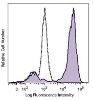

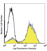
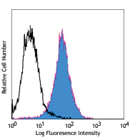
Compare Data Across All Formats
This data display is provided for general comparisons between formats.
Your actual data may vary due to variations in samples, target cells, instruments and their settings, staining conditions, and other factors.
If you need assistance with selecting the best format contact our expert technical support team.
-
Biotin anti-mouse CD206 (MMR)
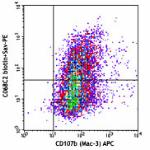
Thioglycollate-elicited Balb/c peritoneal macrophages were s... 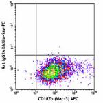
-
Purified anti-mouse CD206 (MMR)
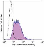
Thioglycollate-elicited BALB/c mouse peritoneal macrophages ... -
FITC anti-mouse CD206 (MMR)
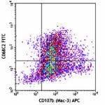
Thioglycollate-elicited Balb/c macrophages were fixed/pemeab... 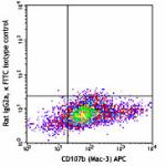
-
PE anti-mouse CD206 (MMR)
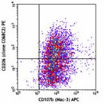
Thioglycollate-elicited BALB/c peritoneal macrophages were s... 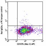
-
APC anti-mouse CD206 (MMR)
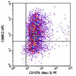
Thioglycollate-elicited BALB/c peritoneal macrophages were s... 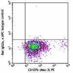
-
Alexa Fluor® 488 anti-mouse CD206 (MMR)
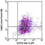
Thioglycollate-elicited BALB/c peritoneal macrophages were s... 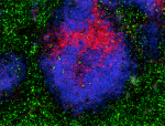
C57BL/6 mouse frozen lymph node section fixed, permeabilized... 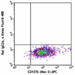
-
Alexa Fluor® 647 anti-mouse CD206 (MMR)
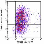
Thioglycollate-elicited BALB/c peritoneal macrophages were s... 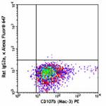
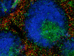
C57BL/6 mouse frozen lymph node section was fixed with 4% pa... -
PerCP/Cyanine5.5 anti-mouse CD206 (MMR)
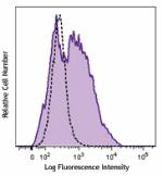
Thioglycollate-elicited BALB/c mouse peritoneal macrophages ... -
PE/Cyanine7 anti-mouse CD206 (MMR)
Thioglycollate-elicited BALB/c mouse peritoneal macrophages ... -
Brilliant Violet 421™ anti-mouse CD206 (MMR)
Thioglycollate-elicited BALB/c mouse peritoneal macrophages ... 
C57BL/6 mouse frozen lymph node section was fixed with 4% pa... 
Confocal image of C57BL/6 mouse thymus sample acquired using... 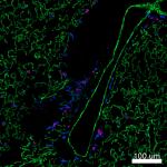
Confocal image of C57BL/6 mouse lung sample acquired using t... 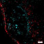
Mice were injected subcutaneously with sheep red blood cells... 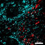
Mice were injected subcutaneously with sheep red blood cells... -
Brilliant Violet 605™ anti-mouse CD206 (MMR)
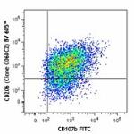
Thioglycollate-elicited Balb/c peritoneal macrophages were f... 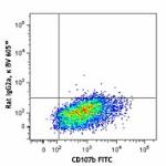
-
Brilliant Violet 650™ anti-mouse CD206 (MMR)
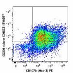
Thioglycollate-elicited BALB/c mouse peritoneal macrophages ... 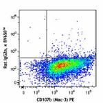
-
Alexa Fluor® 594 anti-mouse CD206 (MMR)
C57BL/6 mouse frozen spleen section was fixed with 4% parafo... -
Brilliant Violet 711™ anti-mouse CD206 (MMR)
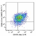
Thioglycollate-elicited Balb/c peritoneal macrophages were f... 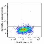
-
Brilliant Violet 785™ anti-mouse CD206 (MMR)
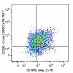
Thioglycollate-elicited Balb/c peritoneal macrophages were f... 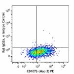
-
PE/Dazzle™ 594 anti-mouse CD206 (MMR)
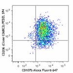
Thioglycollate-elicited Balb/c peritoneal macrophages were f... 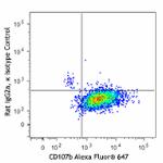
-
Alexa Fluor® 700 anti-mouse CD206 (MMR)
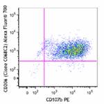
Thioglycollate-elicited BALB/c peritoneal macrophages were f... 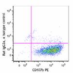
-
Spark YG™ 570 anti-mouse CD206 (MMR)
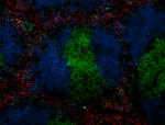
C57BL/6 mouse frozen spleen section was fixed with 4% parafo... -
PE/Cyanine5 anti-mouse CD206 (MMR)
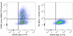
Thioglycollate-elicited BALB/c peritoneal macrophages were f... -
PE/Fire™ 700 anti-mouse CD206 (MMR)
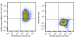
Thioglycollate-elicited BALB/c peritoneal macrophages were s... -
TotalSeq™-B0173 anti-mouse CD206 (MMR)
-
Spark Red™ 718 anti-mouse CD206 (MMR) (Flexi-Fluor™)
-
Spark Blue™ 574 anti-mouse CD206 (MMR) (Flexi-Fluor™)
-
TotalSeq™-C0173 anti-mouse CD206 (MMR) Antibody


Follow Us